Cotton wool spots, causes, symptoms, diagnosis & treatment
Cotton wool spots are believed to occur secondary to ischemia from retinal arteriole obstruction 4. Cotton wool spots is thought to represent nerve fiber layer infarct and pre-capillary arteriolar occlusion. However, factors responsible for focal interruption of axoplasmic flow in the retinal nerve fiber layer may result in similar intra-axonal.

Hypertensive Retinopathy Ento Key
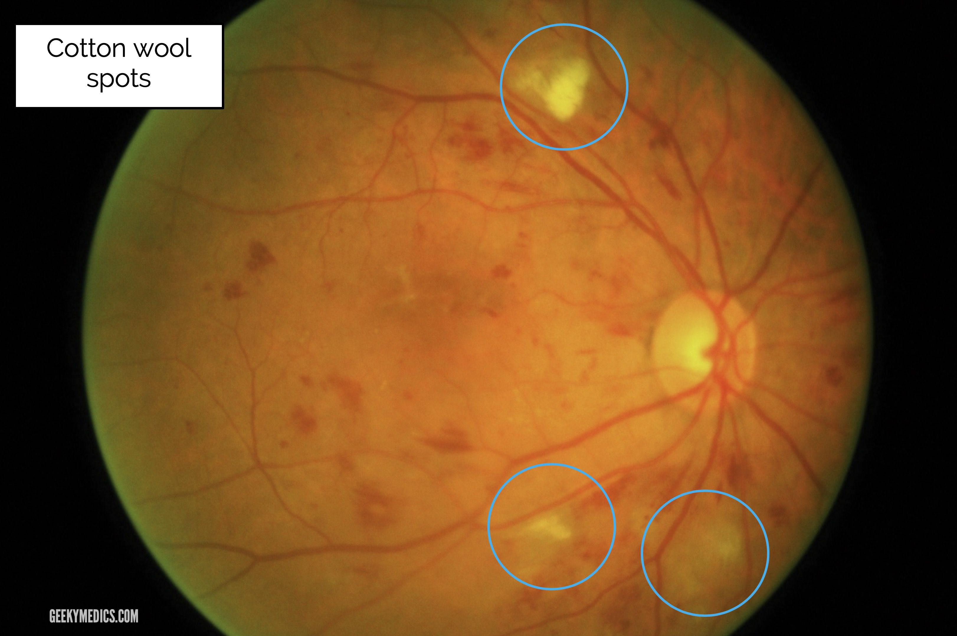
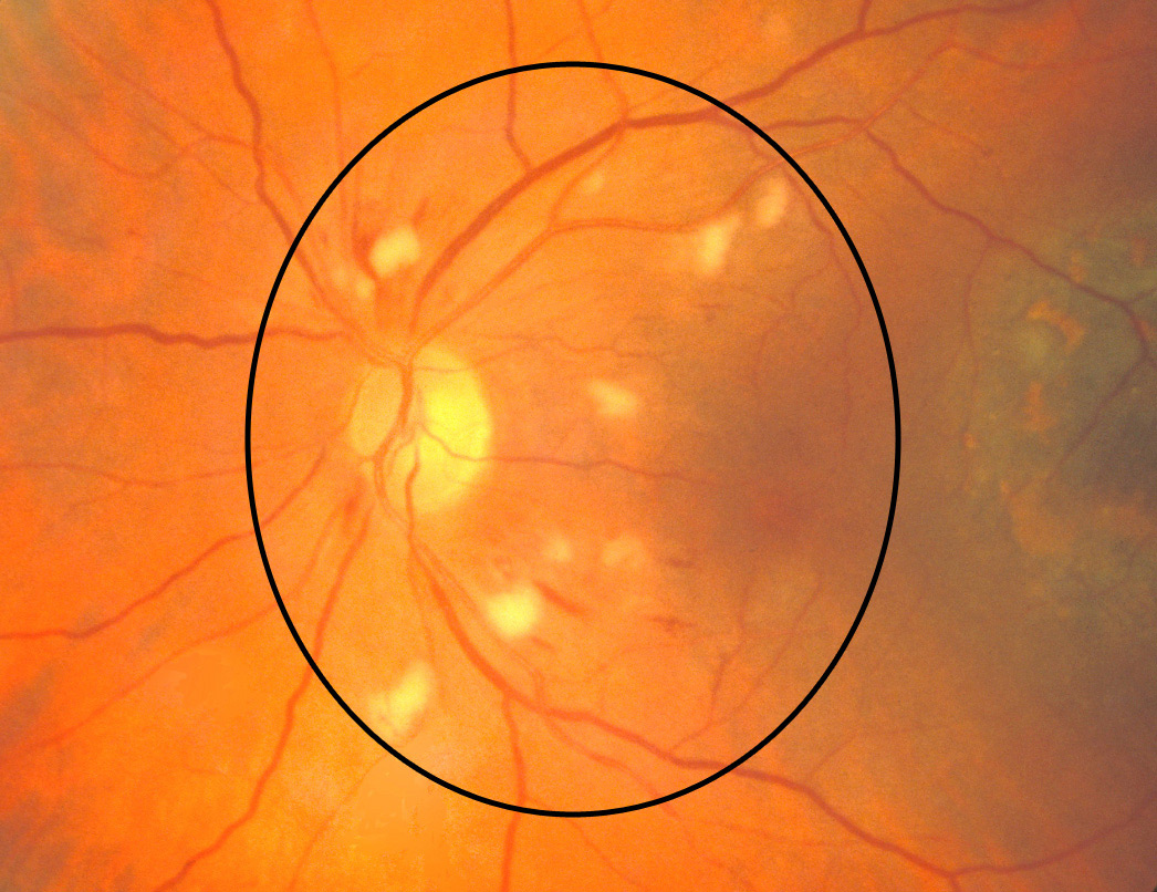
One of these potential retinal findings is the cotton wool spot (CWS). A CWS appears as a white and fluffy superficial lesion 0.1mm to 1.0mm in diameter that obscures the underlying retinal detail. 1,2 This small but important finding can be a marker for potentially life-threatening conditions, making it of great clinical utility.

Solitary cottonwool spot in the right eye ofa patient with PGL who... Download Scientific Diagram
Cotton wool spots are opaque fluffy white patches on the retina of the eye that are considered an abnormal finding during a funduscopic exam (also called an ophthalmoscopic exam). [1] Cotton wool spots are typically a sign of another disease state, most common of which is diabetic retinopathy. [2]

Peripapillary cotton wool spots with hemorrhages were seen in the right... Download Scientific
Cotton Wool Spots : Ophthalmoscopic Abnormalities : The Eyes Have It Cotton Wool Spots What is it? How does it appear? What else looks like it? What to do? What will happen?
Fundus color photographs showing cottonwool spots, exudates, multiple... Download Scientific
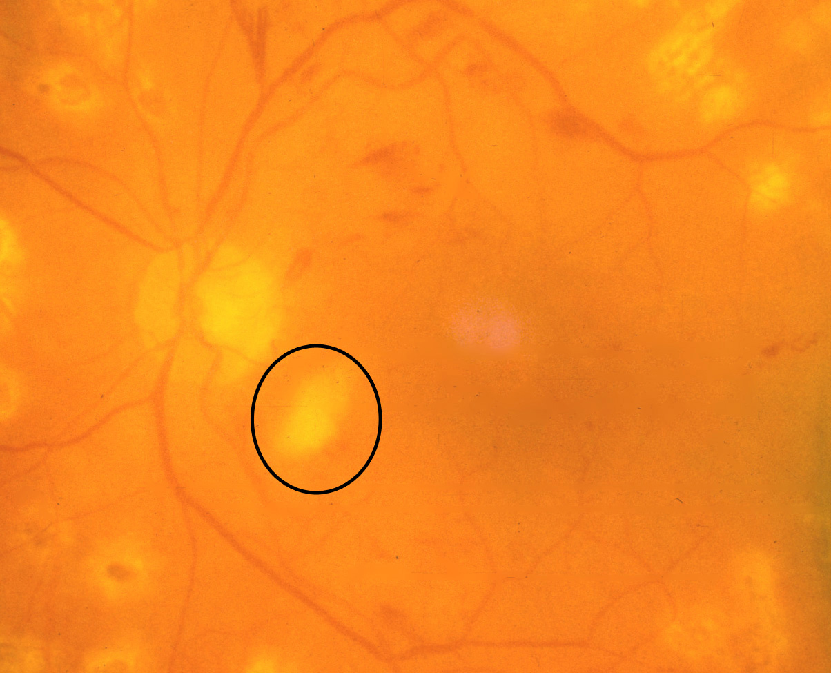
1. "Cotton wool" spots When blood clots prevent nutrients from getting to the retina, the tissue in the retina begins to swell and die. If the doctor examines your eye closely using optical coherence tomography, this area looks white and fluffy like cotton wool (shown in the image above). These spots do not typically affect a person's vision. 2.

Retinal Images BARS
A cotton-wool spot is the name given to a small white spot in the retina that resembles cotton wool (raw cotton). The retina in your eye is like the film inside a camera. The retina "takes the picture" of objects you look at and sends the message to the brain. The retina is a living tissue, which requires blood supplied by tiny vessels.
Fundoscopic Appearances of Retinal Pathologies Geeky Medics
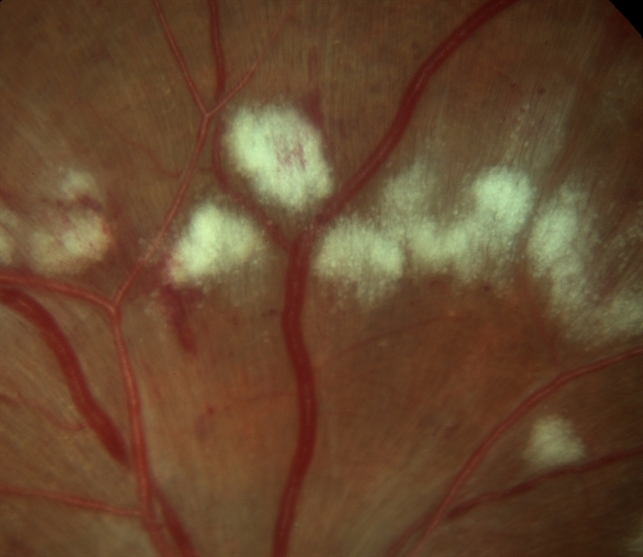
Cotton-wool spots Retinal vasculitis may result in micro-infarcts of the retinal nerve fiber layer that manifests as diffuse, fluffy, cotton-wool like spots in the superficial retinal surface. [1] [12] Systemic vasculitidis such as systemic lupus erythematosus, [36] polyarteritis nodosa, [37] Churg-Strauss syndrome [38] can be associated with.
Cotton wool spots. COMS Grading Scheme
Cotton-wool spots (CWSs) are retinal lesions, most commonly seen as manifestations of diabetes mellitus and systemic hypertension. They are also associated with a number of other etiologies including ischemic, embolic, connective tissue, neoplastic, and infectious, 1, 2 but occasionally no underlying cause can be identified. 2

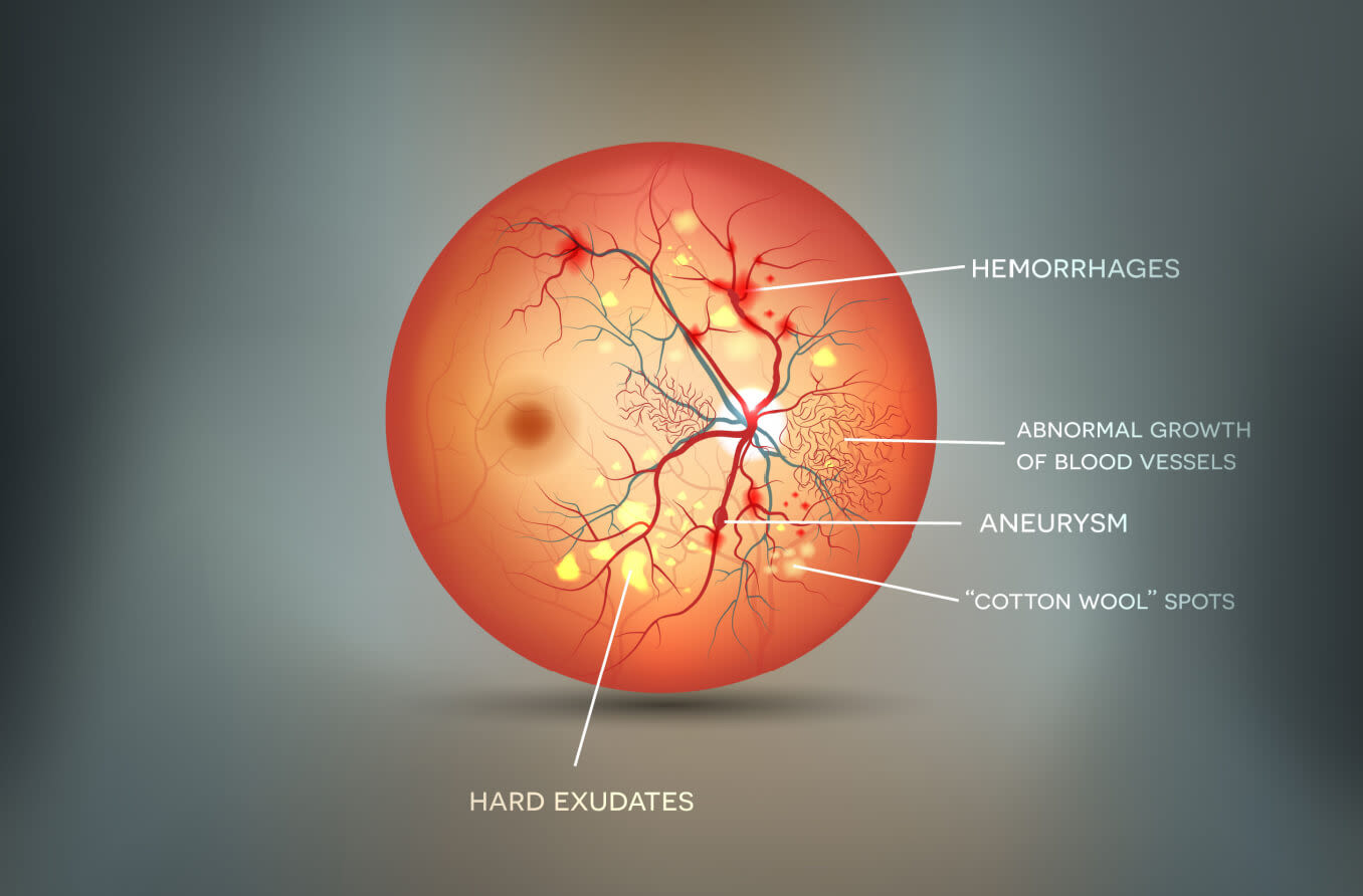
A sample retinal image with cotton wool spots and hemorrhages. Download Scientific Diagram
Retinal cotton-wool spots. In contrast to a vascular occlusion of the precapillary arterial flow, mechanical distortion or traumatic laceration of the nerve fiber layer can also result in the interruption of axoplasmic flow and the development of a cotton-wool spot.
Cotton Wool Spots disease entity and management
Cotton wool spots (CWS) are fluffy white or yellow spots that can appear on the retina. While the spots themselves don't typically cause problems, they often indicate an underlying condition. A CWS can be a cause for concern in an otherwise healthy individual. What causes cotton wool spots?

Cottonwool spots American Academy of Ophthalmology
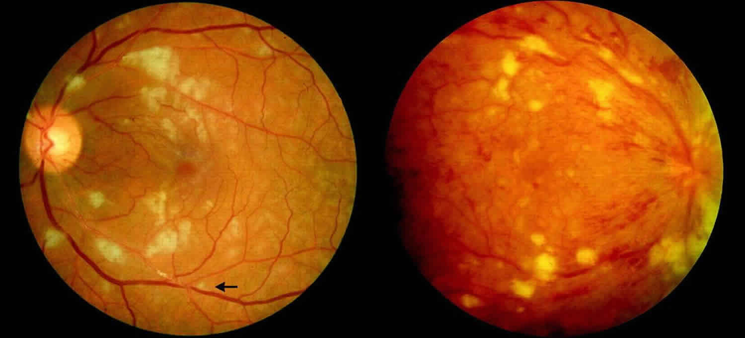
Introduction Hypertension is a risk factor for systemic conditions that can lead to target-organ damage. Specifically, hypertension may lead to multiple adverse effects to the eye that can inevitably cause cause retinopathy, optic neuropathy, and choroidopathy.

CottonWool Spots and Retinal Hemorrhages Clinical Pharmacy and Pharmacology JAMA
Purtscher retinopathy has since been described as a chorioretinopahy associated with indirect trauma, non-ocular injury, associated with a constellation of retinal findings including cotton-wool spots, retinal hemorrhages, optic disc edema, and Purtscher flecken (areas of inner retinal whitening).
Cotton wool spots. COMS Grading Scheme
Axonal myelination in the human central nervous system is a complex, orderly process carried out by oligodendrocyte progenitor cells, which migrate under the influence of neuro-hormonal signals to generate oligodendrocytes that produce myelin.
Cotton Wool Spots Causes and Symptoms
The retinopathy associated with SLE is the most common type of posterior segment finding and the risk of retinal involvement varies with disease control. It may range from 3 percent in well-controlled patients to 29 percent in patients with more active systemic disease. 6,7 The most common retinal manifestation is cotton wool spots ( See Figure.

Why cotton wool spots should not be regarded as retinal nerve fibre layer infarcts British
Cotton-wool spots (CWSs) are common retinal manifestations of many diseases including diabetes mellitus, systemic hypertension, and acquired immunodeficiency syndrome. Clinically they appear as whitish, fluffy patches on the retina and eventually fade with time. In this study, spectral domain optical coherence tomography (SD-OCT) with mapping.

(AB) Fundoscopic examination revealed bilateral cotton wool spots... Download Scientific Diagram
Key Points Manifestations of diabetic retinopathy include microaneurysms, intraretinal hemorrhage, exudates, macular edema, macular ischemia, neovascularization, vitreous hemorrhage, and traction retinal detachment. Symptoms may not develop until late in the disease.